Head joint – Wikipedia


When Head joint the joints between the skull base and the first cervical vertebrae, the Atlas (Atlanto Okipital joint) as well as the joints between Atlas and the second cervical vertebra, the Axis (Atlanto-Axial joints) are referred to. Together with the other cervical spine, these joints cause the mobility of the head in the three room levels: transverse (“turn”), coronal (“tilt”) and sagittal (“nod”). Colloquially, this upper area of the cervical spine is referred to as “neck”. [first]
The upper head joint or Atlantic-Okzipital-Gellek ( ARTICULTE INTLANTOOCCIPITAL ) lies between the two condylene of the head ( Occiput ) and the Pit articular cranialis of the atlas. It is an ellipsoid joint that mainly enables stretching and flexion, i.e. nicking movements (therefore also in English as „Yes“-Joint , German “Yes” joint designated). Sideways’ inclinations of the head are also possible to a lesser extent.
The joint capsule is dorsal (backwards) and ventral (bellyward) through membranes ( Atlantooccipital membrane dorsalis and ventralis ) reinforced. In the area of the dorsal membrane there is a larger hole between the two cervical vertebrae only through this membrane. In this area you can use a cannula into the Subarachnoidal room or its expansion ( Cistern cerebellomedullaris ) To penetrate to carry out a puncture of Cerebrospinalis (brain back fluid, cerebrospinal fluid). You can also destroy the spinal cord there with a pointed object (“neck stitch”). The Membrana Tectoria runs over both head joints in the vertebral canal, below it is the Ligamentum Cruciforme Atlantis.
The Lower head joints or Atlantic-Axial-Gelenke ( Articulatio AtlantoAaxialis ) are formed by Atlas and Axis. There are the following joints:
- Articulatio AtlantoAaxialis Mediana : The vertebral body of the axis is (cranial) upwards by a tap -shaped “tooth” ( Dens axis ) continued, which comes from the Atlas in development. This tooth forms with its Facies articularis anterior In the tooth pit of the atlas ( Pit tooth ) a so-called wheel or tap joint ( Articulatio trochoidea ). Furthermore, the Dens Axis articulates with his Facies articularis posterior with the Ligament transverse Atlantis , which also secures him against reverse movements at the same time. Interestingly, there are bearings of fiber cartilage cells on the surface of the volume, which allow a conclusions to be drawn about a fiery contact with the Dens Axis. The tape lies dorsally of the dens and is on the two Lateral mass of the atlas.
- In the Articulatio atlantoaxialis lateralis stand atlas and axis over the lower and upper joint surfaces of the joint processes ( Articulares process ) in connection.
These joint sections are surrounded by a common joint capsule and fixed by several other ligaments. Around the dens of the axis are predominantly rotating movements like when shaking the head ( „No-joint“ , “No” joint ) executed. The tap joint on the Dens enables 20 ° – 30 ° rotation to each side. About 70% of the head rotation happens in this lower head joint, the rest in the other cervical spine.
The head joints enable a very fine gradation of the movements of the head. Movements in all three room levels are possible by combinating the nod of the upper and rotating movements of the lower head joints.
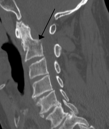
In the event of a break – a break of the tooth of the second cervical vertebrae ( Dens axis ) – or a tear of the ligaments of the Dens axis the extended mark (Medulla Oblongata) and the spinal cord can be cut or squeezed, which leads to a destruction of the breathing and circulatory center. This results in immediate death, comparable to a beheading. In the case of injuries without spontaneous breathing, there is suspicion of a fracture of the Dens axis , so a necessary intubation with caution in the neutral position of the cervical spine must be carried out in order to avoid possible or further damage to extended marks or spinal cord. [2]
A lack of or incomplete training of the Dens axis can be the cause of an atlanto-axial subluxation. This can trigger the same symptoms as with a broken neck.
All alleged “instabilities” of the head joints, which go hand in hand with the ventral atlantodal joint without abnormalities, are unproven claims that have no relevance in conventional medicine and cannot trigger any kind of symptoms. [3] [4] [2] [5] [6]
The explanation for this is that the atlas is the 1st cervical vertebrae, which is designed in a ring and around the Dens axis turns. This lies in the front section, which is why it articulates with the bony front of the atlas arch and forms the front atlantodal joint. The rear joint surface of the Dens axis articulates only with the cross -shaped Ligament transverse Atlantis , which is fixed on the right and left of the atlas, as well as up and down on the back of the back and the 2nd cervical vertebrae ( Bundle longitudinal superior und inferior ). The Dens axis is also hung on its head on the bottom and has two sideways -shaped ligaments ( Corpse. Alare ) that keep him in his position. This ensures that the Dens axis cannot press onto the spinal cord, except for a break or when hanging on the gallows.
The ligaments cannot be seen or assessed on X -ray images, so they are usually not mentioned in an X -ray finding. So there is no dorsal bony atlantodental joint Dens axis the one with that Ligament transverse Atlantis articulated. Rooving or an MRI often provides information about the position and state of the dens and axis, on the basis of which any malformations can also be excluded. In the course of X-ray diagnostics, the DENS with anterior-posterior radiation is carried out, and an MRI of the cervical spine in the event of a striking atlantodal joint. If there is an inconspicuous atlantodal joint – regardless of whether there is an asymmetry to the left or right, since this is a common standard variant without medical relevance – there is no instability of the head joints. [7]
- J. Fanghänel, F. Pera, F. Anderhuber u. (Ed.): Waldeyer anatomy of man . 17. Completely revised edition. de Gruyter, Berlin / New York 2003, ISBN 3-11-016561-9, Chap. 8.2.4 Head joint , S. 640 ff .
- ↑ Definition of the neck in Duden; Retrieved on August 14, 2011
- ↑ a b Distance, atlantodentale. Accessed on June 29, 2019 .
- ↑ Instability of the upper cervical spine. In: Charité Berlin spine therapy. Accessed on June 29, 2019 (German).
- ↑ Sample X -ray images and associated diagnoses. University of Bern, accessed on June 29, 2019 .
- ↑ BNC / Professional Association of Need Surgery Surgery Proctology Pediatric Surgery Vascular Surgery Hand Surgery. (No longer available online) archived from Original am 29. June 2019 ; accessed on June 29, 2019 . Info: The archive link has been used automatically and not yet checked. Please check original and archive link according to the instructions and then remove this note.
- ↑ Deutscher Ärzteverlag GmbH, editorial team Deutsches Ärzteblatt: The cervical spine and cervical market trauma: neurological diagnosis and differential diagnosis. 22. May 1998, accessed on June 29, 2019 .
- ↑ The cervical spine. Accessed on July 1st, 2019 .
Recent Comments