Amoebozoa – Wikipedia

before-content-x4

The Amebozoi I am an important clade of Protozoi Ameboids, understanding the majority of those who move by means of an internal cytoplasmic flow. Their pseudopods are typically smoothed and similar to fingers, called lobopods. Most are unicellular, and are common in the soils and aquatic habitats, with some found, such as symbipons of other organisms, including several pathogens. Amebozoa also include melmic molds, multinucleate or multi -cellular shapes that produce spores and are usually visible to the naked eye.

after-content-x4

Amebozoa vary considerably in terms of size. Many are only 10-20 microns of size, but also include many of the largest protozoa. The famous species Amoeba proteus It can reach 800 microns in length, and in part due to its size it is often studied as a representative cell. Multinucleate Amebe such as Chaos It is Pelomyxa They can be of several millimeters of length, and some muddy molds cover several square meters.

The cell is usually divided into a central granular mass, called endoplasma, and a transparent external layer, called ectoplasma. During the Endoplasma locomotion it flows forward and the ectoplasma flows back along the external part of the cell. Many Amebe move with a front and rear defined, in essence, the cell works as a single pseudopodo. They usually produce numerous transparent projections called subpseudopodes (or certain pseudopods), which have a defined length and are not directly involved in locomotion.

Other Amebozoa can form more indeterminate pseudopods, which are more or less tubular and are largely filled with granular endoplasm. The cellular mass flows into a main pseudopodo, and the others basically portrayed unless you change direction. The subpseudopods are generally absent. In addition to the few naked shapes such as Amoeba and Chaos, the group includes many anebozoa with shell. These can be made up of organic materials, such as in the Arcella genre, or particles collected cemented together, as in that spreads, with a single opening from which pseudopods come out.

The main power mode is phagocytosis: the cell surrounds the particles of potential food, keeping them in these vacuoles where they can be digested. Some Amebe have a rear bulb called Uroide, which they can use to accumulate waste and periodically detach from the cell. When food is scarce, many species can form cysts, who transported from the air, make them know new environments. In muddy molds, these structures are called spores, and form in structures with stem called fruits or sporangi bodies.

Most of the Amebozoa do not have flagelli and, more generally they do not produce structures supported by micro -tubes, except during mitosis. However, the Archamoebae group present Flagelli, and the muddy molds generate many biflagellated gametes. Flagelli are generally anchored by a micro -tubile cone, suggesting a close opistoconti relationship. The mitochondrias typically have branched tubular ridges, but they have been lost by the archiamebe.

Amebozoa are difficult to classify, and the relationships within the group remain confused.

It seems (based on protooms) that form a group of brother of animals and mushrooms, diveting from this line after they had divided from the plants [first] , as illustrated below:

after-content-x4

Plantae

AMOEBOZOA

Fungi

Animalia

Strong similarities between Amebozoi and opistoconti (animals including mushrooms), led to the proposal that they form a clade called Unikonta.

Amebozoi Bosi [ change | Modifica Wikitesto ]

Traditionally all the amebe with lobose pseudopods have been treated together in the lobose class, placed with other ameboids in the Sarcina or Rhizopoda phylum, but these have been considered unnatural groups. Structural and genetic studies have identified the percolzoi and the different architects as independent groups. In the phylogenesis based on RRNA, their representatives were separated from the other Amebe, and it appeared that they diverge close to the basis of the evolution of eukaryotes, as for most of the muddy molds.

However, phylogenetic trees reviewed by Cavalier-Smith and Chao in 1996 [2] They suggested that the remaining loboses form a monofiletic group, and that the architects and micezoa are closely related to it, even if the perolzoi are not. Subsequently they proposed amoebozoa as a phylum major to refer to this supergroup [3] . Studies based on other genes have provided strong support for the unit of this group [4] . Patterson treated most of them together with what they call ” Testate Philosis Amobe ” come ” Ramicristates [5] , on the basis of mitochondrial similarities, but the latter have now been moved to the Cercozoa group.

The lobose subgroup is paraphiletic. Two large classes of lobose have been identified, the tubulinea and flabellinea, but various others remain of uncertain location.

Other amebozoi [ change | Modifica Wikitesto ]

The Archiamebe and Micetozoi have been placed in a conesteen subphylum. This classification receives support from molecular phylogenesis.

Vase microfossils discovered all over the world show that Amebozoi exist from the neopro -nopotoial era. Fossils of the species Melanocyrillium hexodiadema , Palaeoarcella Athanata It is Hemisphereeriella decorated they come from 750 million years rocks. All three fossils have an hemispherical form, inordinated opening and regular recesses, which resemble very modern arcellinids, which are amoeboids with shell. There P. Athanata , in particular, has the same aspect of the existing genus Arcella. [6] [7]

  1. ^ L. EICICHINER, J.A. Pachebat; Glöckner; M. A.Randream; R. SUCGANG; M. RORRIMAN; J. Song; R. Olsen; K. Szafranski; Q. XU;, The genome of the social amoeba Dictyostelium discoideum , in Nature , vol. 435, n. 7038, 2005, pp. 43–57, doi: 10.1038/nature03481 .
  2. ^ T.Cavalier-Smith & E. E. Chao, Molecular phylogeny of the free-living archezoan Trepomonas agile and the nature of the first eukaryote , in Journal of Molecular Evolution , vol. 43, 1996, pp. 551–562, two: 10.1007/BF02202103 .
  3. ^ T.Cavalier-Smith, A revised six-kingdom system of life , in Biological Reviews of the Cambridge Philosophical Society , vol. 73, 1998, pp. 203–266, two: 10.1017/S0006323198005167 .
  4. ^ S. L. Baldauf et al , A kingdom-level phylogeny of eukaryotes based on combined protein data , in Science , vol. 290, 2000, pp. 972–977, two: 10.1126/science.290.5493.972 .
  5. ^ David J. Patterson, The Diversity of Eukaryotes , in American Naturalist , vol. 145, 1999, pp. S96–S124.
  6. ^ Porter, H. Susannah, Meisterfeld, Ralf, and Knoll, H. Andrew, Vase-shaped microfossils from the Neoproterozoic Chuar Group, Grand Canyon: a classification guided by modern testate amoebae , in Journal of Paleontology , vol. 77, n. 3, 2003, pp. 409–429.
  7. ^ ( IN ) M. Susannah Porter, The Proterozoic Fossil Record of Heterotrophic Eukaryotes , in Neoproterozoic Geolobiology and Paleobiology , vol. 27, 2006, pp. 1–21, two: 10,1007/1-4020-5202-2_1 . URL consulted on 11 September 2009 (archived by URL Original on September 25, 2019) .
after-content-x4

Amoebozoa — Wikipedia

before-content-x4

THE amibozo ( AMOEBOZOA ) (Greek amooi signifier ” transformation ) Constitute a large group of simple protozoa, The majority moving by internal cytoplasmic waves [not clear] . Not to be confused with Amiboids (also named rhizopods), which are a subgroup of Amibes, with approximately 200 species of heterotrophic unicellular organisms.

after-content-x4

Their nickname is called lobopod. They can more rarely be equipped with flagelles to ensure their mobility. Most are unicellular, and are often encountered in soils and aquatic environments, where they coexist with other organisms. Several are pathogenic. Amoebozoa can also be multicellular and can produce spores, generally visible to the naked eye.

The size of the Amoebozoa varies a lot. Many are only 10 to 20 μm , but some are the size of the greatest protozoa. The famous species Amoeba proteus can reach 800 μm length [ 3 ] , which makes him a giant in the world of amiboids [ 4 ] , and is often studied as representing a cell (due to its size). When they agglutinate, they can cover very large areas. Some amoebas, belonging to different genres, reach a larger size, for example in genres Gromia, pelomyxa And Chaos .

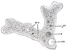
Amibe, as observed by Wilson under a microscope around 1900; Original legend: Amoeba proteus ; An animal made up of a single naked cell x 280 (shot by: Sedgwick and Wilson’s Biology )
n . Nucleus (nucleus); w.v. Water vacuole; c.v. Vacuole contractile ; f.v. Vacuole d’Unfs.

One of the characteristics of the amoebas is that they include one or more nuclei in the same cell, and a contractile vacuole (in) (or pulselle) to maintain their osmotic balance.

The cell is generally divided into a central granular mass, called endoplasm, and an outer layer, called ectoplasm. During locomotion, endoplasmic flows occur at the front and back of the cell. Many Amibes have an anterior side and a posterior side, the cell works as a single pseudopod. In general, they produce numerous projections called subpseudopodia, which are not directly involved in locomotion.

Other Amoebozoa can have multiple indeterminate pseudopods, which are more or less tubular and are mostly filled with endoplasmic pellets. These come together in one of the main pseudopods, and the others retract; They are only used if it changes direction. In addition to a few genres like Amoeba and Chaos, this concerns most of the amoebas that produce a capsule. These capsules can be composed of organic materials, as in Arcella, or collected and glued particles, as in Difflugia, with a single opening by which the pseudopod emerges.

after-content-x4

Most Amoebozoa have no flagella and, more generally, are not supported by a micro -tube network except during mitosis. However, flagelles occur in certain archamoebae, and many genres produce bifellated gametes. Flagelles are generally anchored with a cone of microtubules, which suggests a close relationship with the opisthokonta. Characteristic mitochondria of connection have tubular Cristas, but were lost in archamoebae.

These are non-photosynthetic organisms: they do not produce energy thanks to light. The chlorophyll sometimes observed in their cytoplasms comes from phagocytized microalgae and in the process of assimilation by the amoeba.

The main mode of nutrition is phagocytosis: the cell surrounds potential food particles, then the vacuole closes and absorbs it. Some amoebas have a posterior bulb called a uroid, which can be used to accumulate and evacuate waste, and which periodically detaches from the rest of the cell. When food is rare, most species can form cysts, which can become air and thus be taken to new environments. In myxomycetes, these structures are called spores and form structures called sporocysts.

Amibes are protozoa that occupy almost all compartments of the aquatic environment and wet floors.

The amibe evolves in a strongly aqueous environment and rich in organic matter.
Some do not tolerate hyper saline environments like the sea and develop in water rich in organic matter: marshes, stagnant or polluted waters. It has been found in the cooling waters of thermal power plants and pool waters (poorly treated).

Amibes, like other eukaryotic unicellular organisms, reproduce asexurely by Mitose and Cytocinèse (not to be confused with the binary fission which is the way in which prokaryotes (bacteria) reproduce).

When the amibe is divided by force (cut in half), only the part containing the nucleus will survive and reconstruct a new cell and a cytoplasm. Amibes also do not have a defined form [ 5 ] .

Historically, all pseudopod amibes have been grouped under the class of lobosea, placed with other amiboids in sarcodina or rhizopoda taxa, but these were considered non-natural groups. Genetic and structural studies have shown that Percolozoa and Archamoeba are independent groups. RNA -based phylogeny studies have shown that their representatives were separated from other Amibes, and had to diverge near the basis of the evolution of eukaryotes, as well as most myxomycetes.

However, the trees were revised by Cavalier-Smith and Chao in 1996 and it was suggested that the other lobosans form a monophyletic group, and that Damhamoeba and Mycetozoa are closely linked to it, although the percolozoa are not.

Classification according to BioLib (April 20, 2021) [ 6 ] :

Classification according to World Register of Marine Species (January 20, 2023) [ 7 ] :

We distinguish amoebas free parasitic amibes.

  • Amibes free are capable of evolving perfectly independently in their environment. However, some of them can invest the body of a host if the opportunity is offered to them and trigger a pathology.
  • Parasitic amibes are constantly looking for a host; They survive in the environment but generally develop there little. It happens that some do not trigger pathological syndrome in the host; We will then preferentially speak of commensalism but rarely of symbiosis (the amibe is unicellular).

Some Amibes have pathogenic power for various species, including man.

This is the case of Entamoeba histolytica Responsible for friendship or Amibiase dysentery in tropical circles. The prevalence of this protozoar varies considerably in the different population groups and is generally closely linked to socio-economic conditions.
The highest rates are found in places without sanitary facilities such as toilets, sewers or without access to drinking water.

A particularly dangerous species: Naegleria Fowleri is responsible for a very rare (around 200 cas in the world) but serious pathology: primitive friendship meningo-specimelitis (or meap), almost systematically fatal for humans (~ 97% of mortality within 15 days).

Genre Acanthamoeba like the species Acanthamoeba castellanii is responsible for granulomatous (or EAG) or ocular (keratitis or keratitis)

In addition, we suspect an association between pathogenic amoebas like Acanthamoeba SPP. with pathogenic bacteria such as the genre of Legionella , more particularly Legionella pneumophila , Responsible for legionelloses where the amoeba would play the role of vector and catalyst of secondary infections while retaining, for example, infection households in the bacteria in its cysts.

Immune system in a colony of amoebas [ modifier | Modifier and code ]

Within a colony of amoebas, specialized individuals absorb contaminants (bacteria, toxins). These cells have a storage capacity ten times greater than that of other cells in the colony. They are then rejected at the back as the colony moves. [ 8 ]

Some amoebas show relative resistance to certain biocides (including the active chlorine of swimming pools, under a certain concentration threshold).

Acanthamoeba is more resistant than naegleria (they have a survival duration in water 40 times longer: 40 min against 1 min). 0.5 mg /L of active chlorine (hypochlorous acid) is the minimum necessary in a swimming pool to eliminate amoebas, which implies 0.5 to 1 mg /l of permanently active chlorine. If the hypochlorous acid is activated by 5 to 10% of emerging bromine, the chlorine kills amoeba faster.

Another remarkable characteristic of amoebas is the large size of their genome.

The species Amoeba proteus At 270 billion (10 9 ) base pairs in its genome, and Polychaos doubt (formerly named Amoeba doubtful ) in account 670 billion. The human genome is small by contrast (approximately 2.9 billion bases) [ 9 ] .

Amibes were for the first time described by August Johann Rösel von Rosenhof in 1757 [ ten ] .

For the first naturalists Amoeba The “animal protean” referred to the Greek mythology in which the GOD Proteus could change appearance. The name “Amibe” was then given to these organizations by Bory de Saint-Vincent [ 11 ] , from Greek evangel (fee), signifiant ‘Changement [ twelfth ] ».

DienTamoeba Fragilis was described in 1918 [ 13 ] And has not been recognized as important pathogenic in humans only with difficulty and we still do not know how this amoeba is transmitted (maybe at the same time as the spores of the parasitic worm (oxyer) Enterobius vermicularis) [ 13 ] .

The functional and structural simplicity of Amibes has made it possible to make it a model study and laboratory organization.

They have enabled numerous studies, notably by Balbiani on the location of genetic information in the nucleus (Amibe section).

On other Wikimedia projects:

Related articles [ modifier | Modifier and code ]

Bibliography [ modifier | Modifier and code ]

  1. (in) Thomas Cavalier-Smith, “A revised six-kingdom system of life”, Biological Reviews , Cambridge Philosophical Society, vol.73, n°3, août 1998, p. 232. DOI  10.1111/j.1469-185X.1998.tb00030.x
  2. (in) Sina M. Adl, Alastair G.B. Simpson, Christopher E. Lane, Julius Lukeš, David Bass, Samuel S. Bowser, Matthew W. Brown, Fabien Burki, Micah Dunthorn, Vladimir Hampl, Aaron Heiss, Mona Hoppenrath, Enrique Lara, Line Le Gall, Denis H. Lynn, Hilary McManus, Edward A.D. Mitchell, Sharon E. Mozley-Stanridge, Laura W. Parfrey, Jan Pawlowski, Sonja Rueckert, Laura Shadwick, Conrad L. Schoch, Alexey Smirnov et Frederick W. Spiegel , The Revised Classification of Eukaryotes » , Journal of Eukaryotic Microbiology , vol. 59, n O 5, , p. 429-514 (ISSN  1066-5234 , DOI  10.1111/j.1550-7408.2012.00644.x , read online )
  3. Amoeba proteus  ; Amoebae on the Web, consulté 2009-10-08
  4. Maciver, Sutherland; Isolation of Amoebae ; The Amoebae, consulted 2009-10-08
  5. Amoeba ; ScienceClarified.com
  6. BioLib , accessed April 20, 2021
  7. World Register of Marine Species , accessed January 20, 2023
  8. Émilie Rauscher, Immune system: our defenses are older than we , Science and Life, February 2008, page 77.
  9. Sizing up genomes : Amoeba is king » , on GenomenewsNetwork.org (consulted the ) .
  10. (in) Joseph Leidy , Amoeba proteus » , The American Naturalist , vol. twelfth, n O 4, , p. 235–238 (DOI  10.1086/272082 , read online , consulted the )
  11. Jean-Victor Audouin et al. , Classic Natural History Dictionary , Rey and Gravier, ( read online ) , p. 5
  12. (in) Kimberley McGrath et Stacey Blachford (eds.), Gale Encyclopedia of Science Vol. 1 : Aardvark-Catalyst (2 It is ed.) , Gale Group, , 4136 p. (ISBN  978-0-7876-4370-6 , OCLC  46337140 )
  13. a et b Eugene H. Johnson, Jeffrey J. Windsor, et C. Graham Clark ; Emerging from Obscurity: Biological, Clinical, and Diagnostic Aspects of Dientamoeba fragilis ; Clinical Microbiology Reviews , July 2004, p. 553-570 , Flight. 17, No. 3 0893-8512/04/08.00 $ +0; DOI: 10.1128/CMR.17.3.553-570.2004 ( Résumé ).

external links [ modifier | Modifier and code ]

after-content-x4
after-content-x4