GATTIES BETWEEN – Wikipedia
A wikipedia article, free l’encyclopéi.
The M Aladie or Griscelli syndrome (SG) was described for the first time by Griscelli and Pruniéras in 1978 [ first ] .
SG is a rare hereditary disease characterized by partial albinism associated with other pathologies in the most serious cases. It is a genetic disease with autosomal recessive transmission due to mutations in the genes coding the complexor complex of melanosomes.
Most cases are from the Mediterranean perimeter, including essentially Turkey, however some cases from India were described in 2004 [ 2 ] . Sixty cases were described worldwide at [ 3 ] .
This disease affects both men and women.
Griscelli syndrome is classified into three types [ first ] , [ 4 ] All characterized by the presence of hair with gray reflections, silver (we sometimes speak of golden or dusty reflections), but also by the presence of hypopigmentation at the skin level characterizing albinism [ 5 ] .
In addition, in the type 1 syndrome of neurological damage is observed in type 1 syndrome: convulsions, paralysis, spasticity. The patient will suffer from significant psychomotor delay.
Type 2 is characterized by immunodeficiency (which can also lead to neurological problems). This immunodeficiency is at the origin of various infections at the skin, ENT, respiratory level. It is characterized by leukopenia and thrombocytopenia. In addition, macrophagic activation (hemophagocytic syndrome) can occur, that is to say that there will be phagocytosis of blood elements and release of inflammatory post cytokines. T lymphocytes can also infiltrate the brain.
Hepatosplénomegaly as well as eye problems can also appear.

Diagnostic methods are based on the principle of differential diagnosis: the aim is to gradually eliminate other diseases with symptoms close to those of Griscelli’s disease.
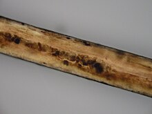
The microscopic study of the hair shows an irregular (clod) distribution of the pigments.
The transmission electron microscopy reveals that melanocytes are overloaded with melanosomes: organelles manufacturing melanin, on the contrary the keratinocytes contain very little melanosomes.
Blood smear reveal the absence of cytoplasmic granules in leukocytes.
Electroencephalograms can highlight neurological problems.
Biopsies of liver and bone marrow are also used for diagnosis.
In addition, for types 1 and 2 there is an antenatal diagnosis it is the removal of chorial villi which will be followed by a sequencing of the genes

The three syndromes of Griscelli meet in the hypopigmentation of the skin.
Skin pigmentation is carried out by specialized melanin producing cells, melanocytes. The melanocytes are at the base of the epidermis, and come from the embryonic ectoderm. The cell body is rounded and has many branched extensions which are part of the Kératinocytes of the upper layer. The extensions, called melanocytic dendrites, allow it to communicate with keratinocytes which synthesize keratin.
The melanocytes make small bags, the melanosomes, containing melanin which is the dark pigment. Each melanocyte supports the protection of 36 keratinocytes thus forming an epidermal unit of melanization. It transfers its melanosomes along the activists’ filaments thanks to a complex transporter composed of three units coded by the Myo5A, RAB27A genes and the MLPH gene [ 2 ] , [ 6 ] .
Type 1 Griscelli syndrome (SG1) [ modifier | Modifier and code ]
The cause of SG1 results from a mutation of the myosin 5A (Myo5A) gene, located on the 15Q221 chromosome [ 7 ] . The SG1 probably corresponds to Elejalde syndrome (in) .
The MyO5A gene codes myosin 5A, a motor protein linking actin and playing a role in the intracellular transport of melanosomes in the dendrites of melanocytes. Thus a mutation of this gene prevents the transport of melanosomes.
Early and severe psychomotor delay is little explained, it is believed that it would be due to the problem of transporting the endoplasmic reticulum in the dendrites of neurons.
Type 2 Griscelli syndrome (SG2) [ modifier | Modifier and code ]
Some nodes and organs (including the brain) are infiltrated by T lymphocytes and macrophages which once activated phagocyte blood cells it is hemophagocytic syndrome. The SG2 results from a mutation of the RAB27A gene (located in the same region as the Myo5A gene) [ 8 ] . On the one hand, it code key effectors in intracellular vesicular transport. So in the event of mutation, the vesicular transport of the melanosomes is modified. On the other hand, the RAB27A protein also regulates the secretion of cytotoxic granules, hence the triggering of hemophagocytic syndrome [ 9 ] [Insufficient source] .
Type 3 Griscelli syndrome (SG3) [ modifier | Modifier and code ]
The SG3 is due to MLPH mutations, a gene coding melanophiline, which forms a protein complex with RAB27A and myosin 5A and participates in the transport of melanosomes in melanocytes.
For SG1, treatment remains symptomatic and limited with taking corticosteroids, antiepileptics, and antipyretics. However, this care cannot generally be able to prevent early death caused by severe neurological involvement.
For the SG2, hemophagocytic syndrome is often fatal and the only curative treatment is bone marrow transplant [ ten ] .
For the SG3, to date no treatment is offered.
The survival prognosis of patients with Griscelli syndrome type 1 and 2 is relatively low, usually, the disease is fatal in 1 to 4 years without treatment. But the disease is more viable for the patient with a type 3 SG, as he has only surface skin problems [ 11 ] .
- (in) Griscelli C, Durandy A, Guy-Grand D, Daguillard F, Herzog C, Prunieras M. « A syndrome associating partial albinism and immunodeficiency » Am J with. 1978; 65: 691-702.
- (in) Martino Ruggieri, Ignacio Pascual Castroviejo, Concezio di Rocco, Neurocutaneous Disorders: Phakomatoses & Hamartoneoplastic Syndromes , Spinger who newyork (ISBN 978-3211213964 ) . p. 418-426
- (in) Fischer A, Virelizier Jl, Arenzana SF et al. « Treatment of four patients with erythrophagocytosis by a combination of epipodophyllotoxin, steroids, intracranial methotrexate and cranial irradiation » Pediatrics 1985; 76: 263-8.
- specific page on the Orphanet site.
- (in) Rajadhyax M, Neti G, Crow Y, Tyagi A. « Neurological presentation of Griscelli syndrome: Obstructive hydrocephalus without hematological abnormalities or organomegaly » Brain Dev. 2007; 29: 247-50.
- http://www.jisppd.com/article.asp?issn=0970-4388;year=2008; .
- (in) Klein C, Philippe N, Le Deist F, Fraitag S, Prost C, Durandy A, Fischer A, Griscelli C. « Partial albinism with immunodeficiency » J Pediatr. 1994; 125: 886-95.
- (in) Ménasché G, Pastural E, Feldmann J, Certain S, Ersoy F, Dupuis S, Wulffraat N, Bianchi D, Fischer A, Le Deist F, de Saint BG. « Mutations in RAB27A cause Griscelli syndrome associated with haemophagocytic syndrome » Nat Genet. 2000; 25: 173-6.
- Hemophagocytic syndrome in another pediatric review.
- (in) Hurvitz H, Gillis R, Klaus R et al. « Kindred with Griscelli disease: spectrum of neurological involvement » Eur J Pediatr. 1993; 152: 402-5.
- http://www.jpad.org.pk/april%20june%202007/11.%20case%20report%20GRiscelli%E2%80%99s%20Syndrome.pdf .
- (in) M Seiberg, ‘ Keratinocyte-melanocyte interactions during melanosome transfer – PubMed » , Pigment cell research , vol. 14, n O 4, , p. 236–242 (ISSN 0893-5785 , PMID 11549105 , DOI 10.1034/j.1600-0749.2001.140402.x , read online , consulted the )
- http://despedara.org/cours_des/sydrome_griscelli_30_11_2007.pdf
Recent Comments