Analysis of blood traces – Wikipedia
L’ analysis of blood traces It is a method of forensic science to analyze the morphology of sketches, patches or bloodstains on the crime scene. The use of blood as proof of the crime, however, is not new in criminology, although there are currently more advanced methods.
The analysis is obtained thanks to the scientific knowledge in concert with other disciplines such as biology, chemistry, mathematics and physics. Following a pre -established analysis plan, it is possible to obtain excellent results regarding scientific investigation.
The analysis of the traces of blood through a series of indicators can help investigators make the investigation framework more clearly:
- Movement of things or people while the crime was taking place;
- Position of things or people during bleeding;
- Movement of things or people after the crime;
- The methods or weapon used for the crime;
- The direction of the sketches;
- The original area of the impact on or with the body;
- The number of shots made to make the crime;
- The sequence of the crime.
The analysis of the traces of blood requires a certain attention and a remarkable preparation, accompanied by the other investigation methods, which are not always taken for granted in the employees of the scientific police.
Analysis indicators [ change | Modifica Wikitesto ]
Once the blood separates from the body, it undergoes all the consequences of the physical laws:
- Severity. He acts on the fluid since his escape. Together with the ballistics it serves to determine the direction of the tracks.
- Viscosity. It corresponds to the amount of internal clutch of the fluid and describes the scrolling resistance.
- Surface tension. It is the strength that determines the shape of the traces. When two fluids are in contact with each other, there are forces that attract molecules each other.
There are several theories on how to classify the traces of blood. Below is a way that is accepted by many based on the mechanism of traces:
Passive training [ change | Modifica Wikitesto ]
The traces of blood are distinguished according to the concomitant factors:
- Passive splashes. The traces are created by the force of gravity;
- Mixed sketches. The traces are created by other traces of blood;
- Flow splashing. The different shape and direction of the track is due to the influence of the force of gravity on the object or on the weapon;
- Patch splashes. A patch is formed when the leakage of blood lasts for a certain period of time.
Active training [ change | Modifica Wikitesto ]
An active trace is formed in conjunction with a force from an external source:
- Low impact splashes. The track is formed by the slightest speed of the impact, e.g. following a fall;
- Medium impact sketches. The track is made up of the intermedia speed of the impact, e.g. a punch in the face;
- High impact splashes. The track is made up of the high speed of the impact, for example, a gunshot;
- Radial splashes. The track is made up of a moving object;
- Sucking Sucks. The track is formed by blood pressure in the artery;
- Specular splashes. The track is made up of a bounce bike towards the source of the impact;
- Areal sketches. The track is formed by the air pressure exerted on the point of leakage.
Meta-Ematic formation [ change | Modifica Wikitesto ]
Based on the contact or transfer of one object on another it is possible to distinguish the type of trace:
- Sketch wipe . The track is formed by an uncontaminated object on an existing track, mutting its consistency.
- Sketch swipe . The track is made up of an object containing traces of blood on another uncontaminated object.
As already mentioned ( see above ) There are other methods used for analysis and different classification models of traces. There is a debate, for example, on the measurement of the speed of the impact. A sub -government commission was established to prepare the most appropriate measures and patterns to obtain a definitive taxonomy.
Contrary to what is written, the “bass-medium-high” terms do not describe the speed of the sketches but rather the amount of energy transferred to the blood source in order to create the track. The speed is measured in meters per second and almost always takes on a certain direction. Often the term strength and energy are mentioned in conjunction with the Anglo -Saxon measurement units. The force is related to speed and mass (n or 1 kg · m · s −2 ). Energy is related to force impressed on an object (j or n · m or kg · m 2 ·s −2 ).
As already mentioned, a debate is underway on how to define the measures. Below is an alternative measurement method. The term “impact” must not be confused with the mechanism with which the track is formed but rather the amount of strength of the track impressed on an object or area:
Low -speed impact [ change | Modifica Wikitesto ]
The low -speed impact occurs when an object, which moves at a speed of less than 1.5 m/s, comes into contact with a source of blood. The size of the track is usually 3 mm diameter higher.
Impact at medium speed [ change | Modifica Wikitesto ]
The medium -speed impact occurs when an object, which moves at a speed between 1.5 m/s and 7.5 m/s, comes into contact with a source of blood. The size of the track is usually between 1 mm and 3 mm in diameter. Among the causes that could determine this type of traces are distinguished the blunt body trauma or the cuts from a blow.
High -speed impact [ change | Modifica Wikitesto ]
The high -speed impact occurs when an object, which moves at a speed greater than 30 m/s, comes into contact with a source of blood. The size of the track is usually less than 1 mm in diameter. Among the causes that could determine this type of traces, explosives and firearms are distinguished.
The experiments on the traces of blood show that a drop of blood tends to form a sphere in the flight trajectory rather than a tear. The sphere shape is the result of the surface tension that stimulates the molecules with each other.
The spherical form is important for the purpose of calculating the impact angle when the sketch meets the surface. This corner is used to determine the point from which the track originated, so -called. “Point of origin”.
A single sketch of blood is not enough to determine the point of origin as it must be evaluated on the basis of the number of traces formed on the opposite part of the surface in order to view a sort of “triangulation”.
Based on the type of shape of the blood track, the investigator can determine the trajectory with which he posed on the surface. This is based on the relationship between the length of the major axis with the minor one and the impact angle.
A very common form of trace is the elliptical one (see figure 1). The French clinician Victor Balthazard discovered the relationship between the width and length of the Elisse that formed the trace and function of the breast of the impact angle. By accurately measuring the track, it was possible to calculate the trajectory of the impact [first] .


Impact [ change | Modifica Wikitesto ]
Due to the three -dimensional appearance of the trajectory, all three impact angles are considered:
,
 ,
, , and
 , and
, and . The easiest angle to calculate is the “range” (
 . The easiest angle to calculate is the “range” (
 ) which is obtained from the vertical of the surface (see figure 2). Another corner that can be easily calculated is the “Alfa” (
) which corresponds to the impact corner that touches the surface of the surface (see figure 2). Finally, the “Beta” corner acts as a pin on the surface (see Figure 2).
Corner calculation [ “> Edit | “> Modifica Wikitesto ]

- = Length of the Elisse (major axis)
- = Elisse width (minor axis)
- = Impact angle


The correlation between these two variables is:

So:

Calculation between two or more corners , , It is [ , , It is “> Edit | , , It is “> Modifica Wikitesto ]


The success of the analysis of the blood traces depends on the diligence of the investor and the ICT competition: the professional software creates an Elisse superimpression on the trail of blood on a scale, therefore it is possible to use a scientific calculation software to complete the measurement of the trajectory. This produces very accurate results that are lent for posthumous investigations and research.
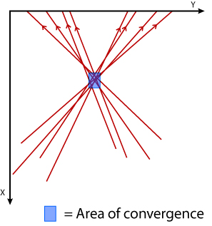
The convergence point is the intersection of two blood trajectories, where the track was formed following the sketch from the opposite side to the impact (see figure 3). To discover the convergence point, the investigator must determine the trajectory of the sketch using the impact angle and the corner opposed to the track, cd. stringing . For analytical purposes, only the view from the top of the trajectory is required (2D), while for an overview you can resort to the convergence area which is the parallelepiped formed by the intersection of more traces of sketches from the opposite side to the impact (see figure 4).
In the past, some analysts designed straight lines along the major axis of a track and compared them on a portion of the wall (convergence area). Rather than seeing it upside down, they preferred to resort to a front point of view. This, however, produced a false point of convergence.

The point of origin is a three -dimensional space where the source of the sketch was at the time of the escape (see figure 5). The space includes the three -dimensional convergence area and the “Z” management, perpendicular to the ground.
To create a point of origin, the agent must be placed in a three -dimensional space and must have a point where only a sketch of blood can be emitted, so that there are many points of origin as the traces analyzed.
As for convergence, the point of origin is calculated through professional software.
In situations where there are heinous crimes, the attention of forensic photography is aimed at traces of blood. The equipment necessary to document the crime scene includes 35 mm machines, digital machines and camera. Often the combination of these tools is used.
There are three types of variable forensic photography depending on the type of lens used:
- Holistic. The large viewing images (28–35 mm range) provide for a good overview.
- Metagrafia. Images taken with normal lenses (45–55 mm range) serve to give a good level of detail of the crime scene. In heinous crimes, metagraphy can grasp only one blood model.
- Close. Images portrayed with slow macro give the best range of details. A medium -speed impact, e.g., can contain hundreds of small tracks (1–3 mm in diameter) many of which require single shots.
Many times the investigator cannot be present directly on the crime scene. So, he has to do all the work in the laboratory on the basics of the images taken by the photographer. An appropriate measurement scale could be the destination accompanied by close images. For all other images, the measurement scale should be parallel and perpendicular to the ground. This is the investigator and whoever works on the images, in order to obtain a neutral perspective on what is being observed.
The bodies are dynamic. In addition to the victim’s somatic movement, the elasticity of the skin and the fragility of the bones is found. Once the strength applies to a body, there must be an equal and opposite reaction to that strength by the attacker. Part of that force will move towards the blood source, even just a millimeter, and will change its trajectory when it will escape the blood. In this way the blood source becomes “contaminated” and misleading for the analysis. The new technologies, therefore, are increasingly oriented on the forecast of the error.
- ^ Balthazard v .; POgelievre r .; Devoence h.; Robert L., Studies of blood drops Projectè, Annals of Legal Medicine , vol. 4, 1939, pp. 265-323.
- Bevel, Tom; Gardner, Ross M. Bloodstain Pattern Analysis With an Introduction to Crimescene Reconstruction , 3rd Ed. CRC Press 2008
- Stuart H. James, Paul Erwin Kish e T. Paulette Sutton, Principles of Bloodstain Pattern Analysis , 3rd, illustrated, revised, Taylor and Francis/CRC Press, 2005, ISBN 0-8493-2014-3. URL consulted on January 30, 2009 .
- Eliopulos L.N. (2008) Murder team. Scientific investigations on the crime scene , Rome, Ed. Mediterranee.
- Hueske E.E., (2002) Shooting Incident Investigation/Reconstruction Training Manual.
- IABPA (International Association of Bloodstain Pattern Analysts). Suggested IABPA Terminology List. Retrieved October 2005 from: https://web.archive.org/web/20051013141146/HTTP://www.iabpa.org/terminology.pdf
- IABPA (International Association of Bloodstain Pattern Analysts). Suggested IABPA Terminology List. Retrieved October 2005 from: https://web.archive.org/web/20051022004009/HTTP://www.iabpa.org/reveduc.pdf
- Stuart J.H., (1999) Eckert, William G. Interpretation of Bloodstain Evidence at Crime Scenes, 2nd Edition, CRC Press.
- Berg S., Villee M. (1993) Biology, 3rd edition. Saunders College Publishing, Fort Worth.
- Sutton, P.T., (1998) Bloodstain Pattern Interpretation, Short Course Manual, University of Tennessee, Memphis TN.
- Vennard J.K. (1982) Elementary fluid mechanics. John Wiley & Sons, New York.
Recent Comments