Lower limb rim – Wikipedia, Free Encyclopedia
From Wikipedia, a free encyclopedia
Lower limb rim (pour. the lower limb of the lower limb ) or pelvic rim It is a set of bones forming a type of ring connecting the free part of the lower (rear) limb with axial skeleton, which includes: spine, skull and chest.
The lower limb rim includes two pelvic bones ( Os Coxae ). Both pelvic bones together with the sacrum and the button form a stable bone ring, called bone pelvis (Pelvis).
The function of the pelvic rim is the combination of lower (rear) limbs – femur – with the lumbar spine. In addition, the pelvis has a protective function towards the epithelial organs and internal pelvis such as urinary tract channels and internal genitals.
Pelvic bones are the largest and widest bones of the human skeleton. Each consists of three parts, which during the childhood are clearly separated by a layer of cartilage, while in an adult they are unity. These are: hip, pubic and sciatic bone. On the outer side of the pelvic bone there are acetabulum – surrounded by the wraps of acetabulum – which are the places of joint femoral joints with the pelvis [first] .
Hip bone [ edit | Edit code ]
The largest pelvic bone consisting of two parts (shaft and plate) is the hip bone. The border between them is marked by the presence of a arc line (Linea Arcuat). The core of the hip bone (Corpus Ossis Ilia) is a thickened part producing the upper stretch of the acetabulum. It is also a place of attachment of part of the straight thigh muscle – from the outside – and the fiber of the internal curtain (on the inside of the bone shaft, as its name suggests) [first] .
Ischium [ edit | Edit code ]
The lower and rear pelvic area forms a sciatic bone (OS ischii). It distinguishes a core that creates a part of the femur as and a branch (Ramus ssis ischii). The sciatica branch is a thin, flattened fragment serving as a field of attachment for the part of the external and internal curtain, some muscle fibers of the adductor and the transverse transverse muscle [first] .
Pubis bone [ edit | Edit code ]
It is a frontal part of the pelvic bone. It is built by a core and two branches – upper and lower. The core is partly formed by the acetabulum – the place of laying the femoral base in the pelvic [first] .
Acetabulum [ edit | Edit code ]
They lie on the outer surfaces of pelvic bones. A spherical shape deep depression shaped by the pubic, hip and sciatic bone. Panewki are places matched to their shapes to the base of the femoral bone, thanks to which they are used to connect to the pelvis with the help of the joint [first] .
Anatomical differences in the construction of pelvis in men and women [ edit | Edit code ]
Sexual differences in pelvic structure can already be observed in the fetus. The pubic joint of the male newborn is visibly higher than the female. However, unambiguous and characteristic sexual differences occur only in a mature person, because during the puberty the pelvis of women is adapted to childbirth.
The female pelvis has a wide side of the hip plates. In men they are arranged more steeply. Women’s cross bone is relatively wider and flatter than male [first] .
-

Fig. 1 Lower limb rim
-
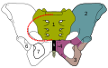
Dig. 2 1- Cross, 2 – hip bone, 3 – sciatic bone, 4 – pubic bone
-


- ↑ a b c d It is f Loaf B. Adam Loaf B. , Richer R. Michael Richer R. , “Human Anatomy” volume I , 1990 .
- human anathomy Edited by Adam Bochenko and Michał Reicher, Warsaw 1990, PZWL


Recent Comments